

Partner: University Hospital Ostrava
Field: Healthcare
 The Ostrava University Hospital is a state-funded organization established by the Ministry of Health of the Czech Republic. Apart from the provision of health care services, its other activities include the development of science and research, as the organisation carries out basic and applied research.
The main objective of the collaboration between the Ostrava University Hospital and the National Centre of Competence in HPC was to deploy and test a tool enabling remote automatic tissue segmentation from patient image data obtained by computed tomography (CT) or magnetic resonance imaging (MRI) on supercomputers operated by the IT4Innovations National Supercomputing Centre. The methods used to enable tissue segmentation were based on deep learning (DL). The objectives were (i) to provide a service based on state-of-the-art algorithms for automatic segmentation of the desired tissues as an AI-based annotation service and (ii) to collect the data after the automatic segmentation and validation by medical doctors, and to provide HPC-based training of new models or enhancement of the existing models through fine-tuning.
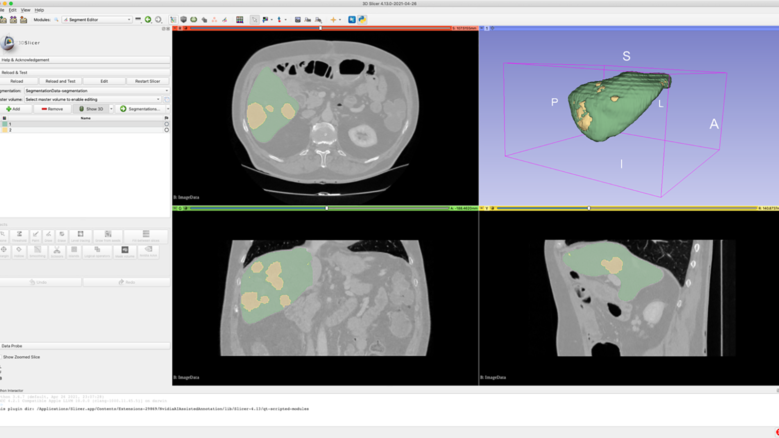
The evaluated concept consists of two main parts. The first one runs at a medical doctor’s site in the hospital, and the other one at IT4Innovations National Supercomputing Center. The part at the hospital is represented by a frontend that mediates the interaction between the doctor and the data. An open-source 3D Slicer was chosen to provide this functionality, and it facilitates the interconnection to a backend part. The backend part provides the computing power and other required functions. It enables deep learning models to be trained for automatic segmentation as well as segmentation of the required tissue from incoming image data. The NVIDIA Clara Train SDK was used to create the backend part.
Analysis of the patient´s medical image data is used to establish or specify the patient´s diagnosis and is performed by medical doctors. When detailed data analysis such as image segmentation is required, e.g. to prepare a patient for surgery, the process is lengthy and demands the physician´s attention given that it is often entirely or largely manual. Consequently, there is a high demand for supporting tools that enable the automation of this radiological examination, especially when the quality evolves with each additional case analysed.
Jan Roman, MD, University Hospital Ostrava
“The use of this toolkit is beneficial for both patients and physicians, as automation allows us to achieve high-quality image reconstructions in a fraction of the time and with minimal effort. In addition, the automated segmentation process can be applied to specific tissues of real interest to the physician, and the resulting model can be further used to plan healthcare tailored to the individual patient.”